Blog

Diagnosis of the difference between coccidiosis and necrotic enteritis symptoms.

Diagnosis of the difference between coccidiosis and necrotic enteritis symptoms

Published on: July 24, 2024
Author: Dr. Ali Salavati

Reading Time:7 minutes

Share on:

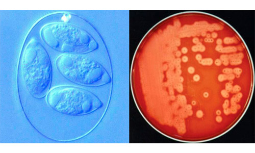
Diagnosing intestinal diseases in poultry is often complex. Given the significant economic losses and the different treatment methods for intestinal diseases, it is essential to know how to differentiate between the distinct symptoms of each disease. Coccidiosis and necrotic enteritis symptoms may appear similar; therefore, recognizing these differences is crucial for making appropriate choices for their control.

In recent years, due to the rise in antibiotic resistance, the use of antibiotics as a preventive tool for certain diseases has been restricted. In the poultry industry, this reality has led to an increase in some diseases, such as necrotic enteritis.

Necrotic enteritis and coccidiosis are two major gastrointestinal diseases in poultry farming that together cause significant economic damage. However, the cause of each disease is different, and consequently, their treatment methods also differ. For this reason, differential diagnosis of these two diseases is extremely important, and special care must be taken to perform it correctly.
بیماری‌های روده‌ای در پرندگان
At the farm level, one of the most important tools for diagnosis is necropsy to assess the injuries present in the intestines. Examining the pathological signs associated with E. maxima, one of the most important species of coccidiosis, is crucial for differentiating it from necrotic enteritis. This is because this species of coccidia closely resembles the lesions caused by Clostridium perfringens, the causative agent of necrotic enteritis.

Examination of the intestinal serosal surface
Accurate diagnosis of intestinal diseases in poultry is often complex. Given the significant economic losses and varying treatment methods for intestinal diseases, it is essential to understand how to differentiate the specific signs of each disease. The symptoms of coccidiosis and necrotic enteritis appear to be similar; therefore, recognizing these differences is crucial in making appropriate choices for controlling them. In recent years, due to the rise in antibiotic resistance, the use of antibiotics as a preventive tool for some diseases has been limited. In the poultry industry, this has led to an increase in certain diseases, such as necrotic enteritis. Necrotic enteritis and coccidiosis are two major gastrointestinal diseases in the poultry industry that cause significant economic losses together. However, the causes of these two diseases are different, and consequently, their treatment methods differ. Therefore, differential diagnosis of these two diseases is very important, and special care should be taken to perform it correctly.
When starting the dissection and removal of the bird’s intestine, it is essential to focus on the duodenum and jejunum junction, especially since this area can create the most ambiguity between the diagnosis of coccidiosis and necrotic enteritis symptoms.
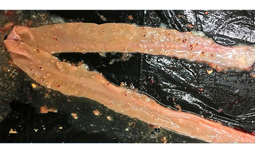
For example, in the case of necrotic enteritis, in addition to the thinning of the wall, the intestine appears bloated due to the presence of gas. In contrast, in coccidiosis, gas production is not present in the disease’s pathogenesis. Additionally, in cases of coccidiosis infection, the intestinal wall is usually thickened.
بیماری‌های روده‌ای در پرندگان
Foci of necrosis with hemorrhage, uneven mucus, and gas in the intestine affected by necrotic enteritis.

Evaluation of specific intestinal lesions
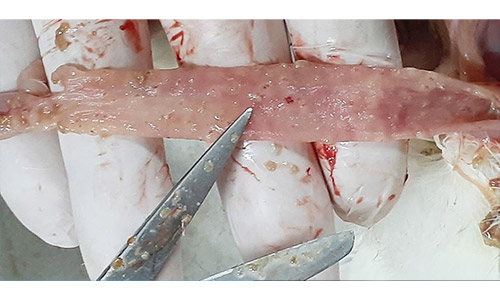
At the level of intestinal mucosal lesions, the Lensing et al. (2010) scoring method is used to assess the severity of Clostridium perfringens increase.
In this assessment system, lesions are categorized based on a severity scale from 0 to 4, ranging from the least to the most occurrence (with 4 corresponding to dead chickens showing symptoms). In this system, the number and size of necrotic lesions in the mucosa are analyzed.
بیماری‌های روده‌ای در پرندگان
Lesions caused by Clostridium perfringens include focal necrosis and superficial petechiae.
The next notable case is petechial lesions, which can be observed on the serosal surface of the intestinal wall. If red spots are seen, it indicates small hemorrhages at a considerable depth in the intestinal wall, which typically represent the foci of oocyst proliferation of Eimeria.
بیماری‌های روده‌ای در پرندگان
Thickening of the intestinal wall and deep petechiae of E. maxima.

Examination of the mucosa and intestinal contents

Upon accessing the interior, the thickness, contents, and condition of the intestinal mucosa should be examined. In cases of necrotic enteritis, in addition to the presence of gas bubbles, a watery or sticky content resulting from poorly digested food in the intestinal lumen is typically observed. If the bird is affected by coccidiosis, the thickness of the intestinal wall increases due to the existing inflammation compared to its normal thickness.
The contents of the intestine may become mucous, creamy, bloody, or orange in color (common in E. maxima), depending on the severity of the damage and the species of Eimeria. In severe cases of necrotic enteritis, the intestinal surface becomes mucous, brown in color, and has a rough appearance (commonly known as “turkey towel”).

Related articles

Contact us

“Please enter your information and a short message. * indicates required fields.”

Diagnosis of the difference between coccidiosis and necrotic enteritis symptoms

Published on: July 24, 2024

Author: NikaVit Team

Reading Time: 7 minutes

Share on:

Diagnosing intestinal diseases in poultry is often complex. Given the significant economic losses and the different treatment methods for intestinal diseases, it is essential to know how to differentiate between the distinct symptoms of each disease. Coccidiosis and necrotic enteritis symptoms may appear similar; therefore, recognizing these differences is crucial for making appropriate choices for their control. In recent years, due to the rise in antibiotic resistance, the use of antibiotics as a preventive tool for certain diseases has been restricted. In the poultry industry, this reality has led to an increase in some diseases, such as necrotic enteritis. Necrotic enteritis and coccidiosis are two major gastrointestinal diseases in poultry farming that together cause significant economic damage. However, the cause of each disease is different, and consequently, their treatment methods also differ. For this reason, differential diagnosis of these two diseases is extremely important, and special care must be taken to perform it correctly.

بیماری‌های روده‌ای در پرندگان
At the farm level, one of the most important tools for diagnosis is necropsy to assess the injuries present in the intestines. Examining the pathological signs associated with E. maxima, one of the most important species of coccidiosis, is crucial for differentiating it from necrotic enteritis. This is because this species of coccidia closely resembles the lesions caused by Clostridium perfringens, the causative agent of necrotic enteritis.

Examination of the intestinal serosal surface

Accurate diagnosis of intestinal diseases in poultry is often complex. Given the significant economic losses and varying treatment methods for intestinal diseases, it is essential to understand how to differentiate the specific signs of each disease. The symptoms of coccidiosis and necrotic enteritis appear to be similar; therefore, recognizing these differences is crucial in making appropriate choices for controlling them. In recent years, due to the rise in antibiotic resistance, the use of antibiotics as a preventive tool for some diseases has been limited. In the poultry industry, this has led to an increase in certain diseases, such as necrotic enteritis. Necrotic enteritis and coccidiosis are two major gastrointestinal diseases in the poultry industry that cause significant economic losses together. However, the causes of these two diseases are different, and consequently, their treatment methods differ. Therefore, differential diagnosis of these two diseases is very important, and special care should be taken to perform it correctly.
When starting the dissection and removal of the bird’s intestine, it is essential to focus on the duodenum and jejunum junction, especially since this area can create the most ambiguity between the diagnosis of coccidiosis and necrotic enteritis symptoms.
For example, in the case of necrotic enteritis, in addition to the thinning of the wall, the intestine appears bloated due to the presence of gas. In contrast, in coccidiosis, gas production is not present in the disease’s pathogenesis. Additionally, in cases of coccidiosis infection, the intestinal wall is usually thickened.
بیماری‌های روده‌ای در پرندگان
Foci of necrosis with hemorrhage, uneven mucus, and gas in the intestine affected by necrotic enteritis.

Evaluation of specific intestinal lesions
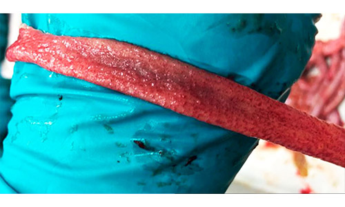
At the level of intestinal mucosal lesions, the Lensing et al. (2010) scoring method is used to assess the severity of Clostridium perfringens increase.
In this assessment system, lesions are categorized based on a severity scale from 0 to 4, ranging from the least to the most occurrence (with 4 corresponding to dead chickens showing symptoms). In this system, the number and size of necrotic lesions in the mucosa are analyzed.
بیماری‌های روده‌ای در پرندگان
Lesions caused by Clostridium perfringens include focal necrosis and superficial petechiae.
The next notable case is petechial lesions, which can be observed on the serosal surface of the intestinal wall. If red spots are seen, it indicates small hemorrhages at a considerable depth in the intestinal wall, which typically represent the foci of oocyst proliferation of Eimeria.
بیماری‌های روده‌ای در پرندگان
Thickening of the intestinal wall and deep petechiae of E. maxima.

Examination of the mucosa and intestinal contents

Upon accessing the interior, the thickness, contents, and condition of the intestinal mucosa should be examined. In cases of necrotic enteritis, in addition to the presence of gas bubbles, a watery or sticky content resulting from poorly digested food in the intestinal lumen is typically observed. If the bird is affected by coccidiosis, the thickness of the intestinal wall increases due to the existing inflammation compared to its normal thickness.
The contents of the intestine may become mucous, creamy, bloody, or orange in color (common in E. maxima), depending on the severity of the damage and the species of Eimeria. In severe cases of necrotic enteritis, the intestinal surface becomes mucous, brown in color, and has a rough appearance (commonly known as “turkey towel”).

Related articles

Contact us

Please enter your information and a short message. * indicates required fields.

Leave a Reply

Your email address will not be published. Required fields are marked *